Blood XX, Skin XY, College
Your word of the day is
par-theh-no-jeh-NET-ik ky-MEER-uh
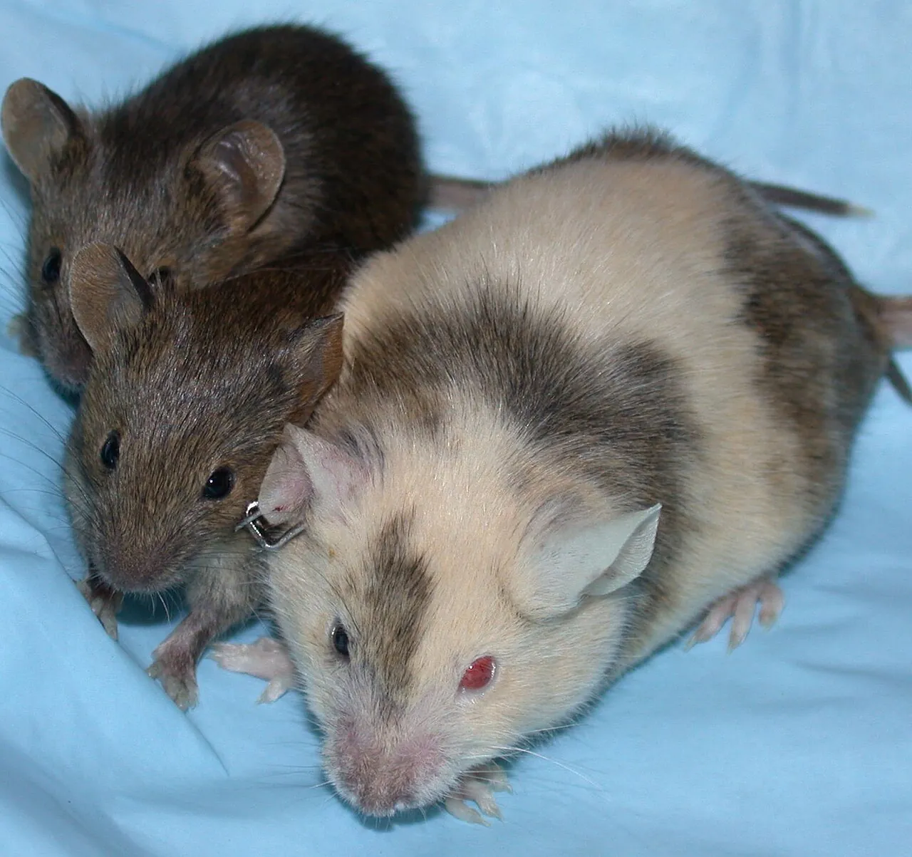
A body made of two cell lines, where one line grew from an egg with no DNA from a father. Also spelled chimera.
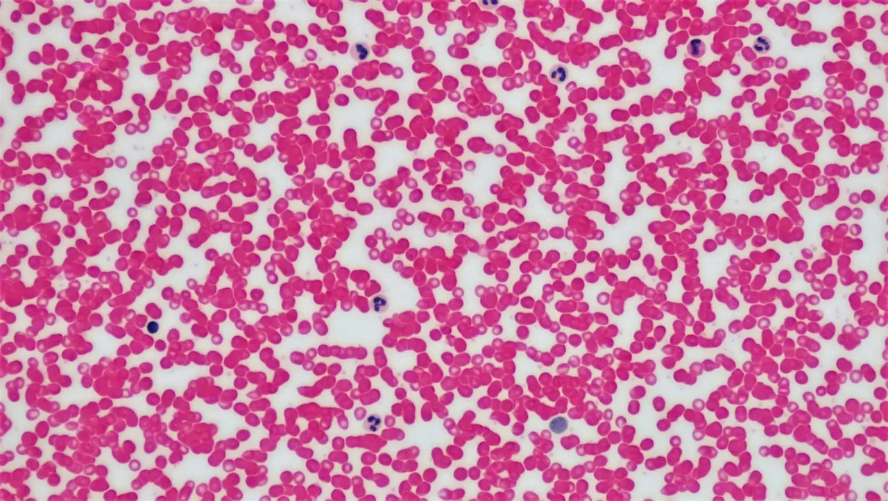
Masunaga and others, 2021, report a new patient whose body holds a mom-only cell line, made of cells carrying only maternal chromosomes.
Before you read the paper by Masunaga and others, make a guess. What percent of their new patient’s white blood cells came from the mom-only line? Write one sentence on why you think so.
Hint
Think about the boy’s blood in the video. Would every patient follow the same pattern?
Read the paper
Masunaga and others, “Parthenogenetic mosaicism: generation via second polar body retention and unmasking of a likely causative PER2 variant for hypersomnia,” Clinical Epigenetics, 2021. The paper is free to read.
Yohei Masunaga’s team reported a 28-year-old woman with parthenogenetic mosaicism, the paper’s term for a mom-only cell line, and compared her case with five earlier reports. Table 1 of the paper lists all six cases, with the boy from the video as Case 1. For each case, the table gives the other cell lines found (the mom-only line was 46,XX in all six) and the estimated share of mom-only cells in each tissue tested.
| Case | Other cell lines | Mom-only share, white blood cells | Mom-only share, second tissue | Proposed mechanism | Gap between tissues (percentage points) |
|---|---|---|---|---|---|
| 1 | 46,XY | More than 98% | Less than 5% (skin fibroblasts) | Parthenogenetic activation and endoreplication | |
| 2 | 46,XY and 69,XXY | About 100% | About 0% (skin fibroblasts) | Unknown | |
| 3 | 46,XY | 0% | 45% (skin fibroblasts) | Parthenogenetic activation and endoreplication | |
| 4 | 45,X | 84% | 56% (saliva) | Parthenogenetic activation and endoreplication | |
| 5 | 47,XXX | Not reported | 42% (amniotic fluid cells) | Parthenogenetic activation and endoreplication | |
| 6 | 46,XX | 70% | 67% (saliva) | Second polar body retention and endoreplication |
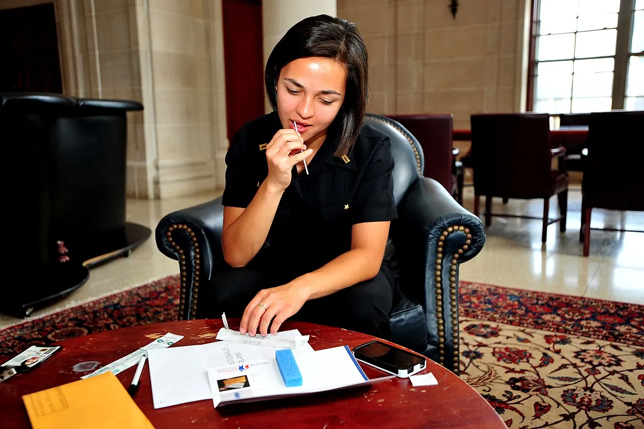
Table 1 gives the mom-only share in two tissues for five of the six cases, and its last column is blank for your answers.
For Case 1 in Table 1, the gap between tissues is more than ____ percentage points.
See the table
| Case | Other cell lines | Mom-only share, white blood cells | Mom-only share, second tissue | Proposed mechanism | Gap between tissues (percentage points) |
|---|---|---|---|---|---|
| 1 | 46,XY | More than 98% | Less than 5% (skin fibroblasts) | Parthenogenetic activation and endoreplication | |
| 2 | 46,XY and 69,XXY | About 100% | About 0% (skin fibroblasts) | Unknown | |
| 3 | 46,XY | 0% | 45% (skin fibroblasts) | Parthenogenetic activation and endoreplication | |
| 4 | 45,X | 84% | 56% (saliva) | Parthenogenetic activation and endoreplication | |
| 5 | 47,XXX | Not reported | 42% (amniotic fluid cells) | Parthenogenetic activation and endoreplication | |
| 6 | 46,XX | 70% | 67% (saliva) | Second polar body retention and endoreplication |
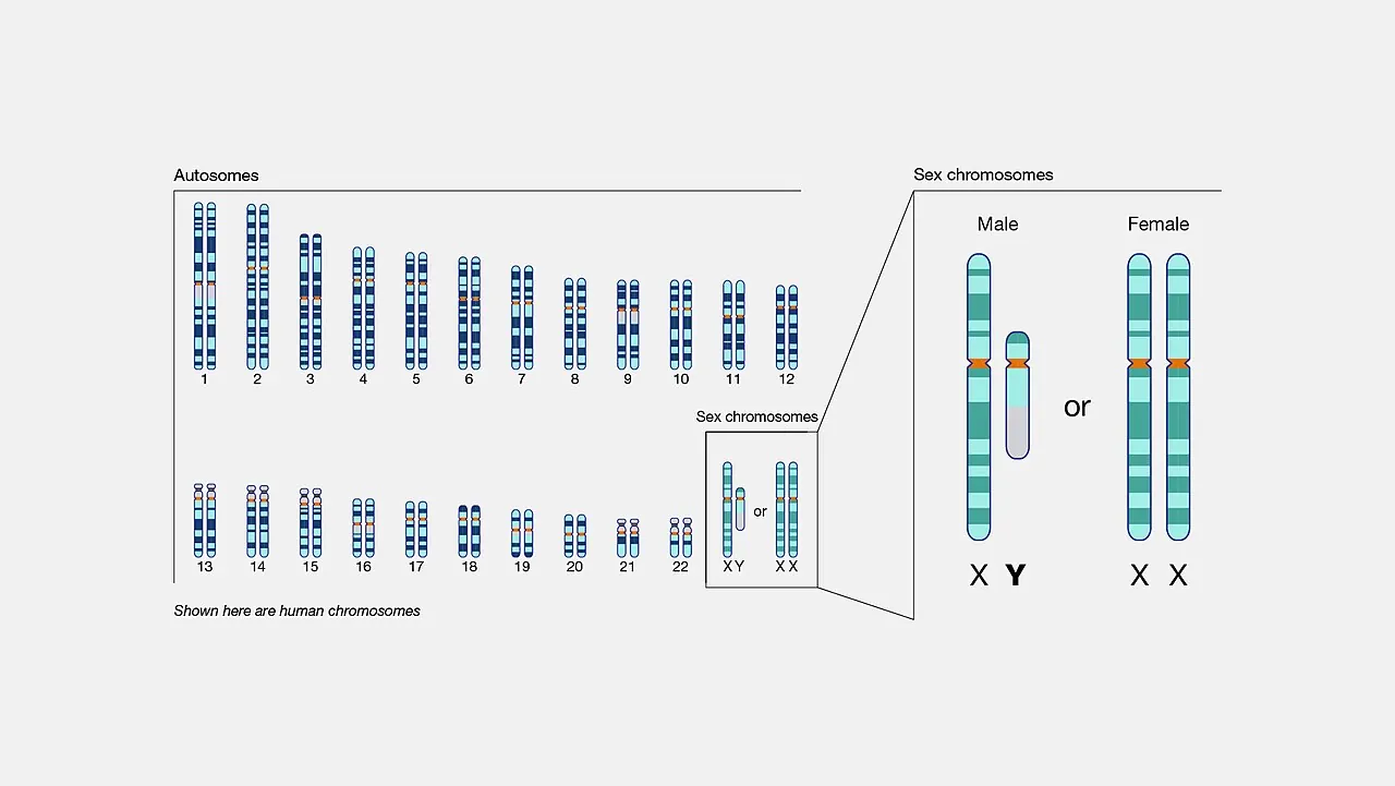
For Case 2 in Table 1, the gap between tissues is about ____ percentage points.
See the table
| Case | Other cell lines | Mom-only share, white blood cells | Mom-only share, second tissue | Proposed mechanism | Gap between tissues (percentage points) |
|---|---|---|---|---|---|
| 1 | 46,XY | More than 98% | Less than 5% (skin fibroblasts) | Parthenogenetic activation and endoreplication | |
| 2 | 46,XY and 69,XXY | About 100% | About 0% (skin fibroblasts) | Unknown | |
| 3 | 46,XY | 0% | 45% (skin fibroblasts) | Parthenogenetic activation and endoreplication | |
| 4 | 45,X | 84% | 56% (saliva) | Parthenogenetic activation and endoreplication | |
| 5 | 47,XXX | Not reported | 42% (amniotic fluid cells) | Parthenogenetic activation and endoreplication | |
| 6 | 46,XX | 70% | 67% (saliva) | Second polar body retention and endoreplication |
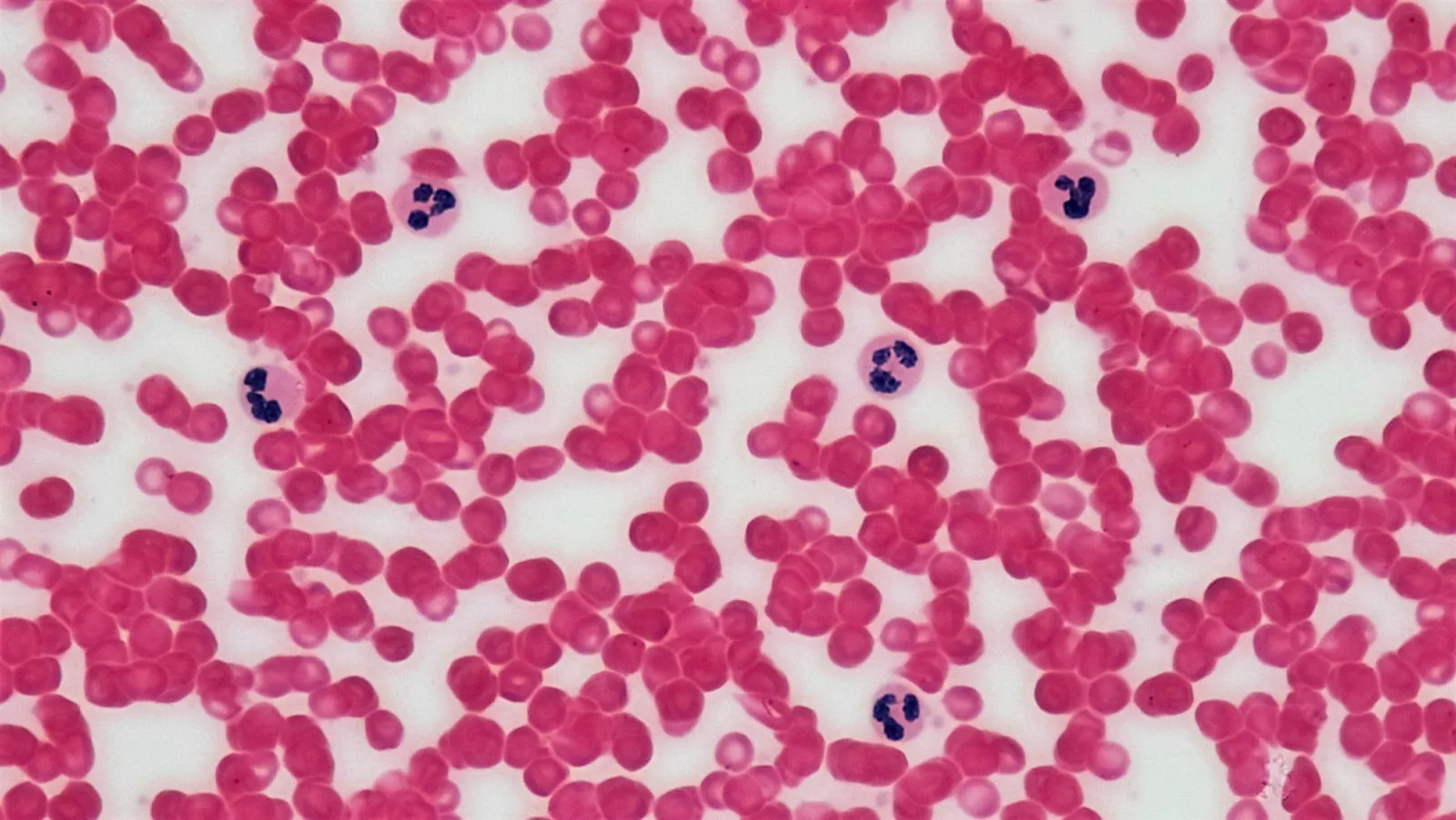
For Case 6, the authors propose that the egg kept its second polar body, which holds sister copies of the egg’s own chromosomes. In Figure 1 and the Results section, regions next to the centromeres carried one version from mom. Some regions farther out carried two different versions from mom.
How does crossing over in meiosis I explain the Case 6 pattern of one maternal version next to the centromeres and two farther out?
See the table
| Case | Other cell lines | Mom-only share, white blood cells | Mom-only share, second tissue | Proposed mechanism | Gap between tissues (percentage points) |
|---|---|---|---|---|---|
| 1 | 46,XY | More than 98% | Less than 5% (skin fibroblasts) | Parthenogenetic activation and endoreplication | |
| 2 | 46,XY and 69,XXY | About 100% | About 0% (skin fibroblasts) | Unknown | |
| 3 | 46,XY | 0% | 45% (skin fibroblasts) | Parthenogenetic activation and endoreplication | |
| 4 | 45,X | 84% | 56% (saliva) | Parthenogenetic activation and endoreplication | |
| 5 | 47,XXX | Not reported | 42% (amniotic fluid cells) | Parthenogenetic activation and endoreplication | |
| 6 | 46,XX | 70% | 67% (saliva) | Second polar body retention and endoreplication |
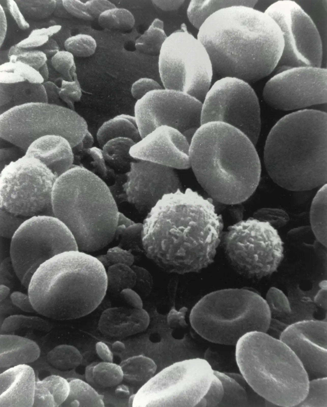
Why does one maternal version next to the centromeres in Case 6 point to one egg instead of two merged embryos?
See the table
| Case | Other cell lines | Mom-only share, white blood cells | Mom-only share, second tissue | Proposed mechanism | Gap between tissues (percentage points) |
|---|---|---|---|---|---|
| 1 | 46,XY | More than 98% | Less than 5% (skin fibroblasts) | Parthenogenetic activation and endoreplication | |
| 2 | 46,XY and 69,XXY | About 100% | About 0% (skin fibroblasts) | Unknown | |
| 3 | 46,XY | 0% | 45% (skin fibroblasts) | Parthenogenetic activation and endoreplication | |
| 4 | 45,X | 84% | 56% (saliva) | Parthenogenetic activation and endoreplication | |
| 5 | 47,XXX | Not reported | 42% (amniotic fluid cells) | Parthenogenetic activation and endoreplication | |
| 6 | 46,XX | 70% | 67% (saliva) | Second polar body retention and endoreplication |
The Case 6 patient has idiopathic hypersomnia, a sleep disorder. Exome sequencing found a rare variant in PER2, a circadian clock gene, in 55 of 66 sequencing reads from her and in 25 of 52 reads from her mother, who has no sleep symptoms.
In the Case 6 patient, ____% of PER2 reads carried the variant.
See the table
| Case | Other cell lines | Mom-only share, white blood cells | Mom-only share, second tissue | Proposed mechanism | Gap between tissues (percentage points) |
|---|---|---|---|---|---|
| 1 | 46,XY | More than 98% | Less than 5% (skin fibroblasts) | Parthenogenetic activation and endoreplication | |
| 2 | 46,XY and 69,XXY | About 100% | About 0% (skin fibroblasts) | Unknown | |
| 3 | 46,XY | 0% | 45% (skin fibroblasts) | Parthenogenetic activation and endoreplication | |
| 4 | 45,X | 84% | 56% (saliva) | Parthenogenetic activation and endoreplication | |
| 5 | 47,XXX | Not reported | 42% (amniotic fluid cells) | Parthenogenetic activation and endoreplication | |
| 6 | 46,XX | 70% | 67% (saliva) | Second polar body retention and endoreplication |
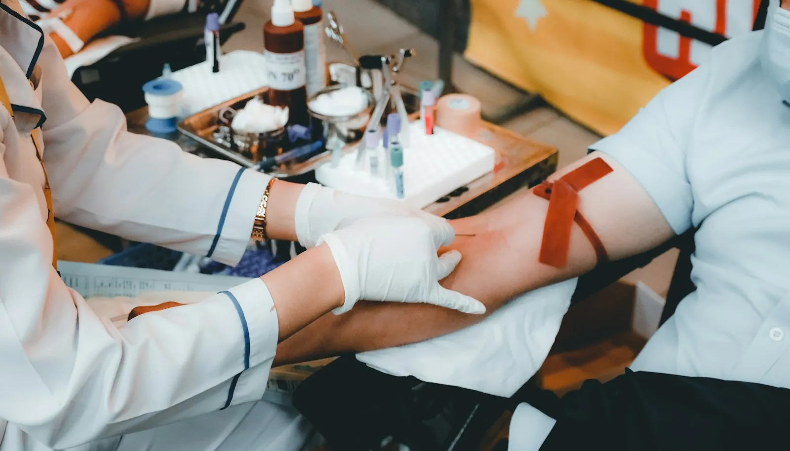
Exome sequencing found the PER2 variant in 25 of 52 reads from the mother of the Case 6 patient.
In the mother of the Case 6 patient, ____% of PER2 reads carried the variant.
See the table
| Case | Other cell lines | Mom-only share, white blood cells | Mom-only share, second tissue | Proposed mechanism | Gap between tissues (percentage points) |
|---|---|---|---|---|---|
| 1 | 46,XY | More than 98% | Less than 5% (skin fibroblasts) | Parthenogenetic activation and endoreplication | |
| 2 | 46,XY and 69,XXY | About 100% | About 0% (skin fibroblasts) | Unknown | |
| 3 | 46,XY | 0% | 45% (skin fibroblasts) | Parthenogenetic activation and endoreplication | |
| 4 | 45,X | 84% | 56% (saliva) | Parthenogenetic activation and endoreplication | |
| 5 | 47,XXX | Not reported | 42% (amniotic fluid cells) | Parthenogenetic activation and endoreplication | |
| 6 | 46,XX | 70% | 67% (saliva) | Second polar body retention and endoreplication |
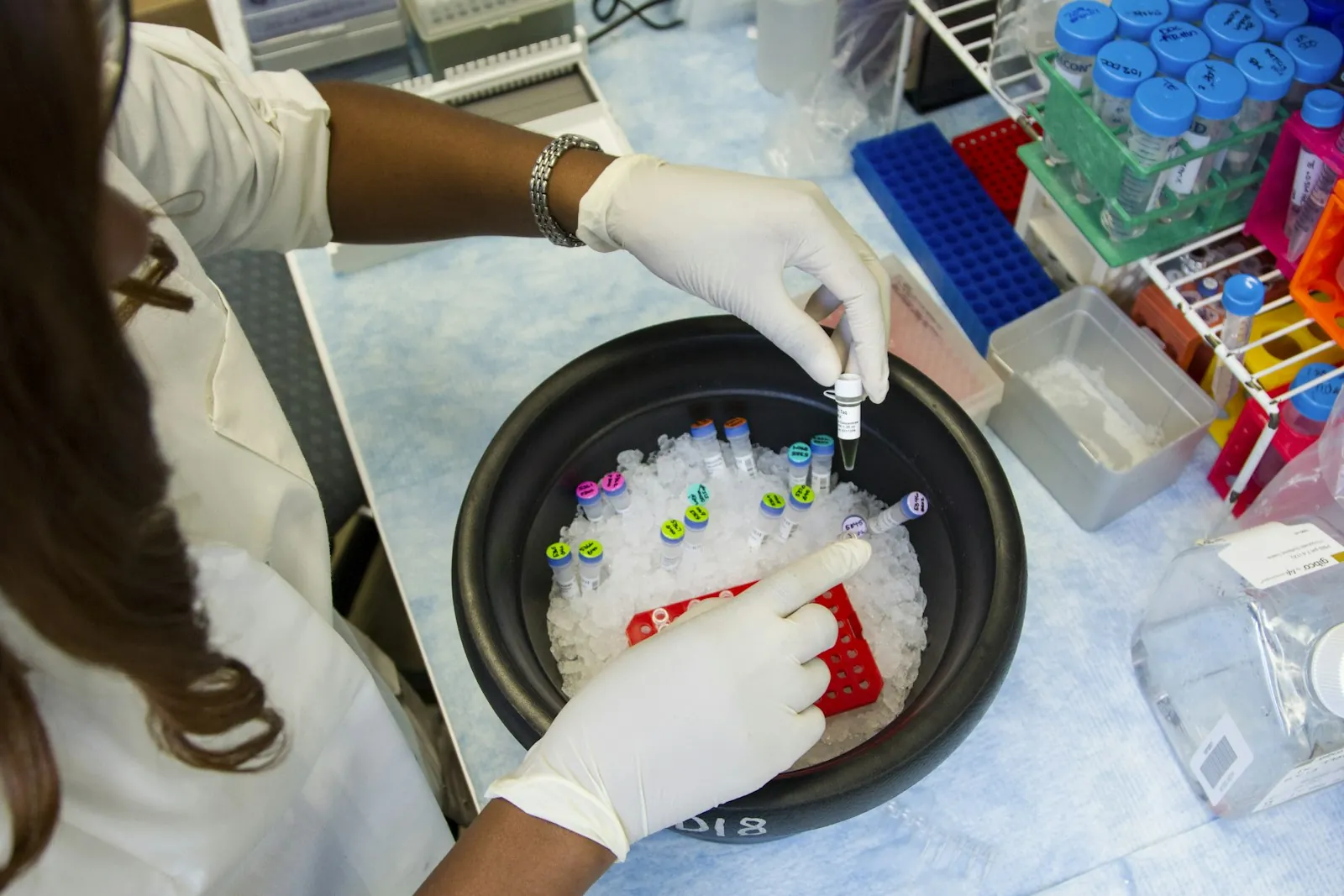
The PER2 reads from the Case 6 patient come from a mix of mom-only cells and two-parent cells.
If 70% of the Case 6 patient’s sequenced cells are mom-only cells with two variant copies of PER2 and each remaining cell has one, the predicted share of PER2 reads with the variant is ____%.
See the table
| Case | Other cell lines | Mom-only share, white blood cells | Mom-only share, second tissue | Proposed mechanism | Gap between tissues (percentage points) |
|---|---|---|---|---|---|
| 1 | 46,XY | More than 98% | Less than 5% (skin fibroblasts) | Parthenogenetic activation and endoreplication | |
| 2 | 46,XY and 69,XXY | About 100% | About 0% (skin fibroblasts) | Unknown | |
| 3 | 46,XY | 0% | 45% (skin fibroblasts) | Parthenogenetic activation and endoreplication | |
| 4 | 45,X | 84% | 56% (saliva) | Parthenogenetic activation and endoreplication | |
| 5 | 47,XXX | Not reported | 42% (amniotic fluid cells) | Parthenogenetic activation and endoreplication | |
| 6 | 46,XX | 70% | 67% (saliva) | Second polar body retention and endoreplication |
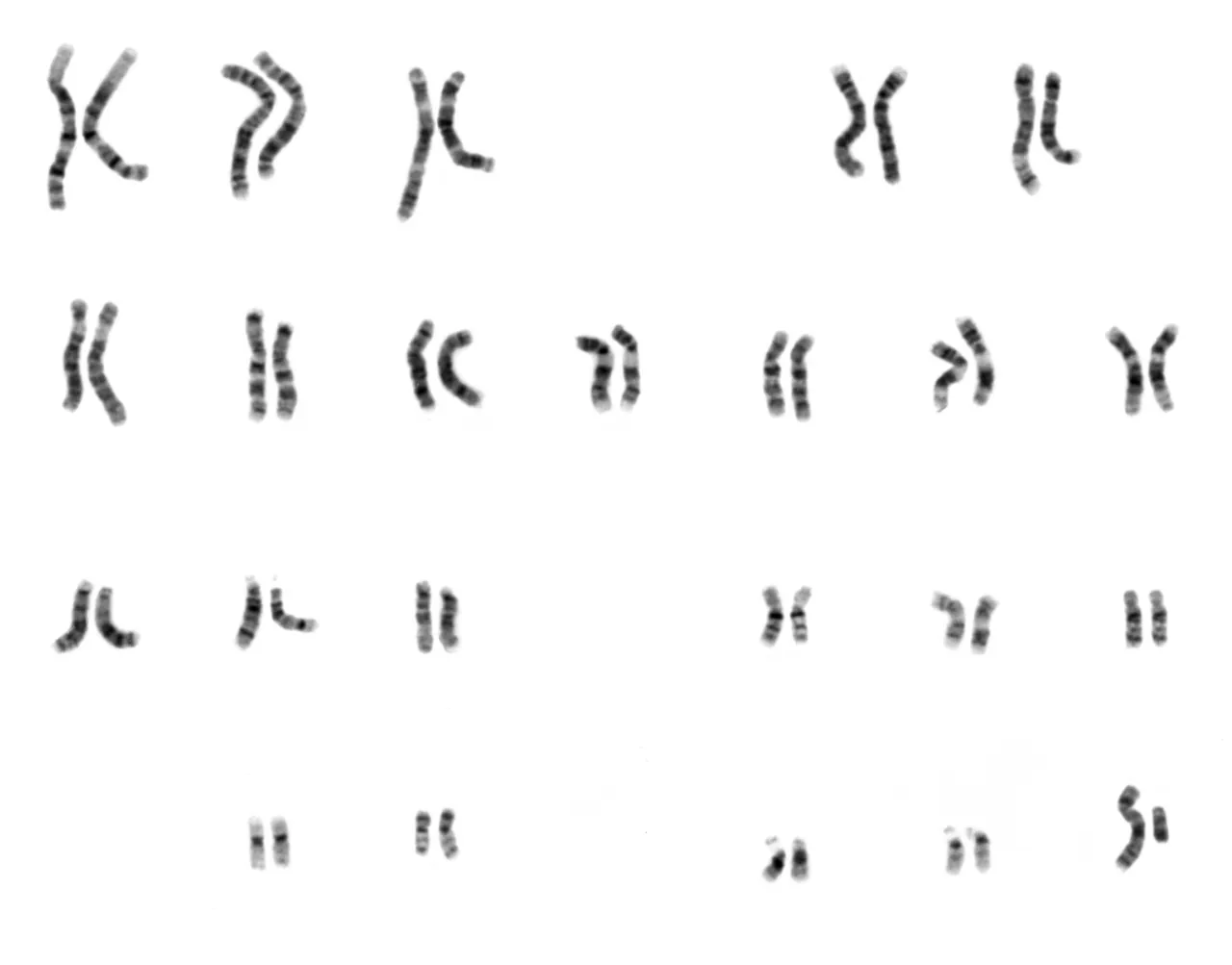
The Case 6 patient’s mother carries one copy of a recessive PER2 variant and has no sleep symptoms.
How can a mom-only cell line give the Case 6 patient two copies of a recessive PER2 variant that her mother carries once?
See the table
| Case | Other cell lines | Mom-only share, white blood cells | Mom-only share, second tissue | Proposed mechanism | Gap between tissues (percentage points) |
|---|---|---|---|---|---|
| 1 | 46,XY | More than 98% | Less than 5% (skin fibroblasts) | Parthenogenetic activation and endoreplication | |
| 2 | 46,XY and 69,XXY | About 100% | About 0% (skin fibroblasts) | Unknown | |
| 3 | 46,XY | 0% | 45% (skin fibroblasts) | Parthenogenetic activation and endoreplication | |
| 4 | 45,X | 84% | 56% (saliva) | Parthenogenetic activation and endoreplication | |
| 5 | 47,XXX | Not reported | 42% (amniotic fluid cells) | Parthenogenetic activation and endoreplication | |
| 6 | 46,XX | 70% | 67% (saliva) | Second polar body retention and endoreplication |
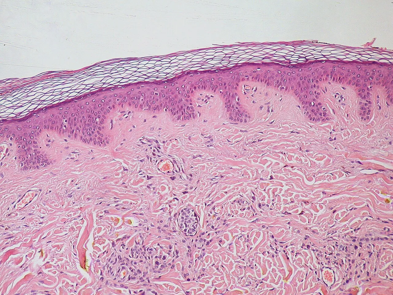
Masunaga and others note a bias in how doctors find people with a mom-only cell line. Genetic tests usually start with blood, and a mom-only line barely present in white blood cells would go unidentified.
When should a genetics clinic test a second tissue, such as skin or saliva, for a mom-only cell line?
See the table
| Case | Other cell lines | Mom-only share, white blood cells | Mom-only share, second tissue | Proposed mechanism | Gap between tissues (percentage points) |
|---|---|---|---|---|---|
| 1 | 46,XY | More than 98% | Less than 5% (skin fibroblasts) | Parthenogenetic activation and endoreplication | |
| 2 | 46,XY and 69,XXY | About 100% | About 0% (skin fibroblasts) | Unknown | |
| 3 | 46,XY | 0% | 45% (skin fibroblasts) | Parthenogenetic activation and endoreplication | |
| 4 | 45,X | 84% | 56% (saliva) | Parthenogenetic activation and endoreplication | |
| 5 | 47,XXX | Not reported | 42% (amniotic fluid cells) | Parthenogenetic activation and endoreplication | |
| 6 | 46,XX | 70% | 67% (saliva) | Second polar body retention and endoreplication |
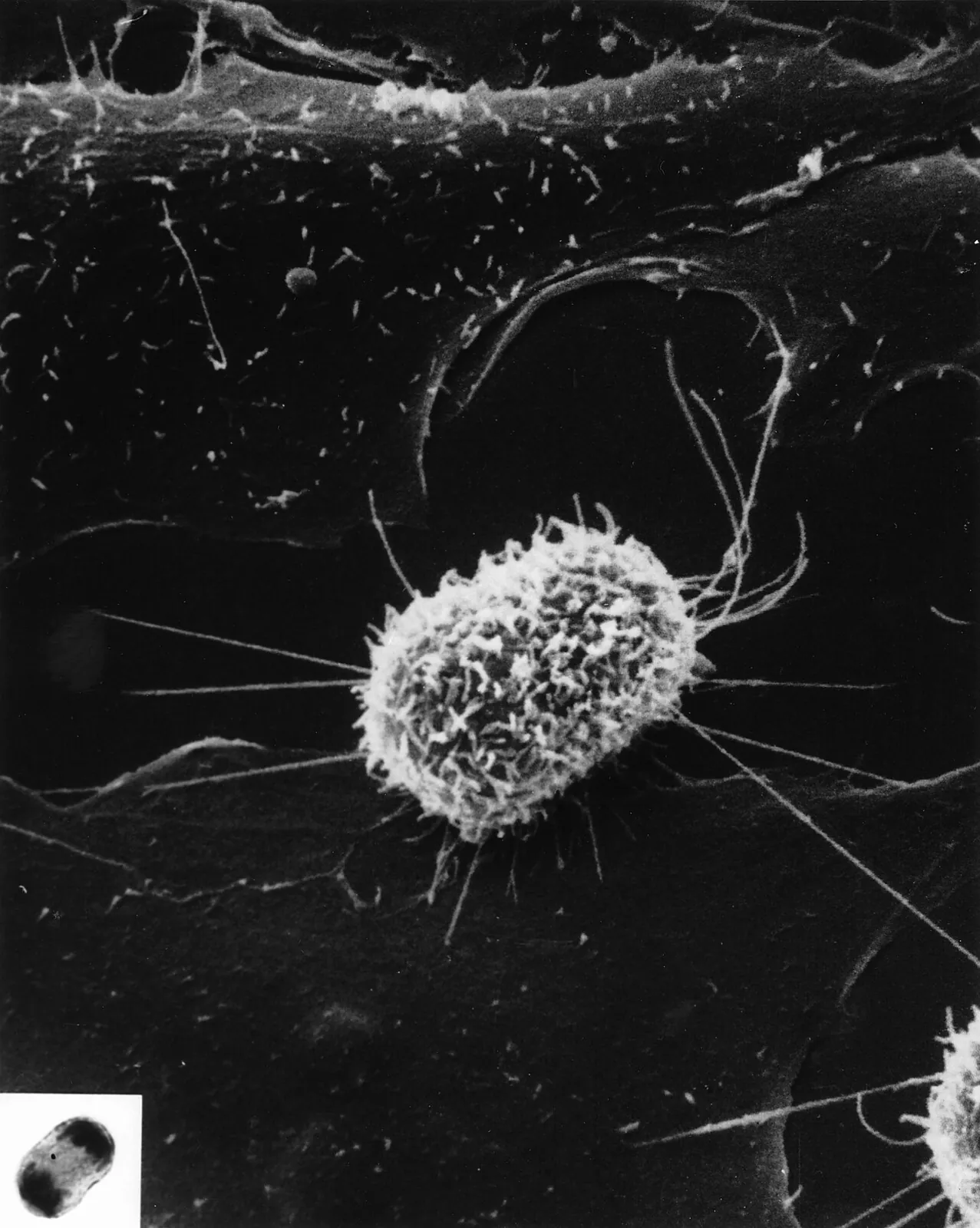
How could testing blood first skew what doctors know about mom-only cell lines?
See the table
| Case | Other cell lines | Mom-only share, white blood cells | Mom-only share, second tissue | Proposed mechanism | Gap between tissues (percentage points) |
|---|---|---|---|---|---|
| 1 | 46,XY | More than 98% | Less than 5% (skin fibroblasts) | Parthenogenetic activation and endoreplication | |
| 2 | 46,XY and 69,XXY | About 100% | About 0% (skin fibroblasts) | Unknown | |
| 3 | 46,XY | 0% | 45% (skin fibroblasts) | Parthenogenetic activation and endoreplication | |
| 4 | 45,X | 84% | 56% (saliva) | Parthenogenetic activation and endoreplication | |
| 5 | 47,XXX | Not reported | 42% (amniotic fluid cells) | Parthenogenetic activation and endoreplication | |
| 6 | 46,XX | 70% | 67% (saliva) | Second polar body retention and endoreplication |
Table 1 of Masunaga and others, 2021, lists all six known cases of a mom-only cell line.
What does Table 1 of Masunaga and others, 2021, show about the six known cases of a mom-only cell line?
Which question can’t Table 1 of Masunaga and others, 2021, answer?
Lesson complete
Today’s word
parthenogenetic chimaera
par-theh-no-jeh-NET-ik ky-MEER-uh
An individual with two cell lines, one of which developed from an egg without fertilization and carries only maternal chromosomes. Also spelled chimera.
Look at these again
You got every question right the first time.
Sources
- Strain and others, Nature Genetics, 1995 (free abstract)
- Yamazawa and others, Journal of Medical Genetics, 2010
- Masunaga and others, Clinical Epigenetics, 2021
- “Scientists: Boy came from egg that didn’t wait for sperm,” Tampa Bay Times, September 30, 1995
- MedlinePlus Genetics: 46,XX testicular difference of sex development, Klinefelter syndrome, Turner syndrome, genomic imprinting and uniparental disomy
- Vision and Change core concepts and competencies, as summarized in Brownell and others, CBE Life Sciences Education, 2014
- Word origins from Dictionary.com: chimera, chimaera, chimerical, genetic, chromosome, chromo-, chromatic, -some, ribosome
- Mark Cartwright, “Chimera of Arezzo,” World History Encyclopedia
- Spanish and French meanings from Wiktionary: quimera, chimère